Black spots often make us worry when we observe them in the mouth. We tend to search a lot about them thinking it to be oral cancer.
But, do you know, not all black spots tend to change into oral cancer? Moreover, it’s very rare to see a black spot turning into oral cancer.
There are very few lesions in the mouth that tend to change into oral cancer from a black spot.
So first of all, there is no need to panic. We provide a step-by-step guide to approaching a dentist and finding out the actual cause of the black spot.
Point to remember:
Take the first step to a better Oral health!
Get tips on Oral health and discover ways to improve your Dental health. Sign up today
Usually, oral cancer presents itself as a growth or an ulcer and never as a black spot. The only lesion that presents as a black spot is Melanoma, which is very rare in the mouth.
what are the reasons of black spots in the mouth?
There are numerous reasons which present as black spots in the mouth. Let us discuss it one by one.
Oral melanotic macule

Oral melanotic macule on lip
Oral melanotic macules (OMM) are harmless freckles in your mouth. They are asymptomatic and do not cause any harm to your body.
The OMM is often seen on lips and cheeks. They represent excess melanin deposition in those areas compared to their surroundings.
The OMMs usually do not turn into malignancy and are inert lesions and tend to be inert throughout their life.
Nevus

Nevus on the chin
In simple words, it is a mole similar to the moles that we see on the skin. The only difference is that they appear inside the mouth.
The nevi are well-demarcated, small to moderately big in size. They usually do not require any treatment and can be diagnosed by simple oral examination.
Nevus is a collection of melanocytes. In simple words, it is a collection of cells that produce melanin. They appear blue to black in color and are harmless.
Point to remember:
Repeated irritation on the nevus may tend to develop oral cancer, but it is very rare.
Trauma (Hematoma / Purpura / Petechiae)
Trauma in the oral cavity may cause bleeding in the tissues. As the bleeding accumulates in the tissues, hemoglobin in the blood discolors the tissue surface.
The main reason for this is that Hemoglobin contains iron which gets deposited in the tissues during trauma. As a result, you will find black pigmentation on the skin or mucosa.
The pigmentation from the trauma is a temporary presentation and will heal with time. They are usually harmless and will not develop into oral cancer.
Eruption cyst in children

Eruption cyst
The main reason for the black spots on gums in children could be the eruption cyst that we commonly see.
Children usually have a lot of teeth an erupting state. During a tooth eruption (Usually milk teeth), we usually see a boil-like structure on the gums.
The boil contains blood and is often painful. The blood in the boil is the main reason why we see black pigmentation in that region.
As previously, mentioned, the iron in the blood discolors the boil and gives a black dot presentation.
These black spots on the gums in children are usually harmless and will subside once the teeth erupt.
Amalgam tattoo
An amalgam tattoo is a form of pigmentation seen in the mouth near metal fillings or restorations. The mercury and its corrosion products leach from the filling as days pass.
The corrosion products get accumulated within the skin or mucosa beside the metal filling. As a result, you might see dark grey to black pigmentation on the cheeks and gums near the mercury filling.
The pigmentation is usually harmless until it starts reacting with the body’s immune system to produce the lichenoid reaction. There is no evidence of lichenoid reactions turning into oral cancer in the literature.
Post-inflammatory pigmentation

Post-inflammatory pigmentation is an interesting condition where we see pigmentation during the healing process of mucosa or skin in the mouth.
During this phase, we find multiple pigmented areas in the mouth, which are preceded by ulcers or abraded areas due to various conditions. Often, the pigmentation is a sign of healing and indicates positive results in the mouth.
The pigmentation is the result of increased melanin deposition from stimulated melanocytes.
We observe post-inflammatory pigmentation in the following conditions:
Anemic stomatitis

Anemic stomatitis is a condition where the mouth becomes inflamed secondary to Anemic condition. Patients suffering from anemic stomatitis present with multiple areas of burning sensation and atrophy of mucosa.
These areas heal by themselves within a span of 1 to 2 weeks and re-appear in new areas. During the process of healing, we find increased pigmentation in these areas that may mimic cancer-like presentation. But these black spots or areas are in no way related to oral cancer.
Moreover, they represent areas of increased melanin deposition.
Xerostomia

In xerostomia, the patient experiences dryness of the mouth. It is a condition where he or she might experience decreased salivary production in the mouth.
In xerostomia, due to dryness, the tissues like the cheek and tongue become dry and produce irritation while moving. As a result of friction, the skin in these areas becomes traumatized and produces a burning sensation. Moreover, these areas heal with pigmentation.
The pigmentations seen in Xerostomia are temporary and might disappear in a few days after the healing of tissues.
Smoker’s melanosis
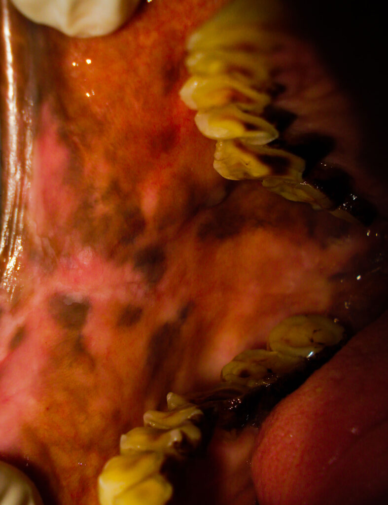
Smokers’ melanosis is an interesting condition often seen in chronic smokers or chain smokers. People with smoking habits often experience large areas of dark pigmentation in their mouths.
One reason could be the excess heat generated within the mouth, which stimulates the melanocytes in the tissue to produce excess melanin.
The second reason could be the deposition of tar and excess dust which are inhaled during smoking. These deposits get impregnated into the superficial tissue causing black pigmentation over the mucosa.
The common areas of pigmentation are the palate and cheeks. You can often find pigmentation on the lips.
Pigmentation in a smoker’s palate often turns out to be dangerous. But the pigmentation itself might not turn into cancer. But, the dust from the smoke entrapped in tissue might cause irritation and might turn into malignancy in patients who are more susceptible to cancer.
Fixed drug eruptions

People who are allergic to any drug will develop Fixed drug eruptions (FDE) if they take the same drug accidentally. In simple words, FDE is a condition where the patient might develop severe lip pigmentations and multiple ulcers in their mouth, after the intake of the allergic drug.
It is a form of anaphylactic reaction that produces gross destruction in the mouth. During this condition, the patient suffers from multiple ulcerations and pigmentations in various regions of the oral cavity.
The pigmentations are well-defined with definite borders and spread across various regions or oral cavities and even on the skin.
Black Spot On Gums Near Wisdom Teeth
A black spot on the gums near wisdom teeth is often an indication of a condition called Pericoronitis.
In pericoronitis, the gums that cover the half-erupted wisdom tooth gets inflamed and swollen. Pus accumulation between the gums and the wisdom teeth is often seen.
As a result, you might see some dark pigmentation over the gums in the region. But it is in no way related to malignant lesions or cancer.
Also read:
What to do next, when I find a black spot in my mouth?
I think by now you understood something about the black spots in the mouth. But what to do next, if i find a black spot in my mouth?
As already discussed, black spots turning into oral cancer is a rare phenomenon. Hence the first thing you should do is “DO NOT PANIC, BE CALM!”
In case of doubt, the first thing to do is to visit a dentist.
Visit a dentist
Once you make an appointment, visit your dentist and ask all the questions that boggle in your mind. Get the right answer for every question you ask.
Have a complete oral examination done. In case your dentist might also feel suspicious about the condition, let him book an appointment with an oral medicine expert.
Ask for an Oral medicine expert consultation
Once a consultation is done with the Oral medicine expert, let him deliver his final diagnosis. In case of any suspicion, he might ask for a blood examination. Further, he might also advise a biopsy.
Get biopsy done
In case your Oral medicine expert advises a biopsy, do not hesitate for the biopsy, kindly cooperate with the dentist and have the biopsy done.
If the biopsy report is negative (Which usually occurs 99% of the time), feel happy about that, and do not forget to thank your dentists. (I think I am too optimistic. Of course, I am, and you too should be !).
If you like the post, do share it with your friends.
- Is oral hygiene compulsory for kids? - January 13, 2023
- How to Choose the Best Dental Crown for Your Smile? - December 8, 2022
- Who needs antibiotics before dental work? What is antibiotic prophylaxis? - October 31, 2022